Juniper Publishers : Very Large, Rapidly Growing Myoma during Second Trimester of Pregnancy - Outcome
JUNIPER PUBLISHERS- JOURNAL OF GYNECOLOGY AND WOMEN’S HEALTH
Journal of Gynecology and Women’s Health-Juniper Publishers
Authored by Angel Danchev Yordanov*
Abstract
Intruduction: The leyomyoma is the most common gynaecological tumor. Uterine myomas during pregnancy occur in 2 to 10% of all cases. One tenth of those may lead to complications of the pregnancy. According to most ultrasounds studies myomas remain the same size or become smaller during pregnancy.
Case: This case report is about 34 year old woman, nullipara, in 14th week of pregnancy. Through ultrasound observation was found subserous pediculated myoma node with diameter of 20cm. When the pregnancy was found, the myoma was not. A median laparotomy with myomectomy was performed without any complications. On the 38th week of pregnancy a healthy, 3400g, full-term baby was delivered by cesarean section.
Conclusion: Though rarely, rapidly growing and very large myomas can be an indication for miomectomy during the second trimester of pregnancy. Laparotomic myomectomy should be preferred over laparoscopic myomectomy in cases of very large myomas, due to prolonged operative time, inapplicability of in-bag morcellation and related risks of dissemination of a leiomyoma or leiomyosarcoma.
Keywords: Very large myoma; Pregnancy; Myomectomy; Rapidly growing myoma
Introduction
The leiomyoma is the most common benign gynaecological tumour [1]. Uterine myomas during pregnancy occur in 2 to 10% of all cases. One tenth of those may lead to complications of the pregnancy. According to most ultrasounds studies myomas remain the same size or become smaller during pregnancy [2-4]. Estrogen and progesterone were thought to play a major role in the development and growth of myomas, but recently this theory has been questioned.
Usually myomectomy is avoided during pregnancy, but in some situations it is indicated, despite the risk of hemorrhagic complications that may require hysterectomy due to the increased vascularity [5-7]. Laparotomic myomectomy during pregnancy has been reported as a safe approach more than a hundred years ago [8,9]. The most common indications for myomectomy during pregnancy are acute pelvic pain that is not responsive to medical therapy, signs of red infarction or torsion of pedunculated myomas, and abdominal discomfort due to a large or rapidly growing myomas [7,10].
Clinical Case
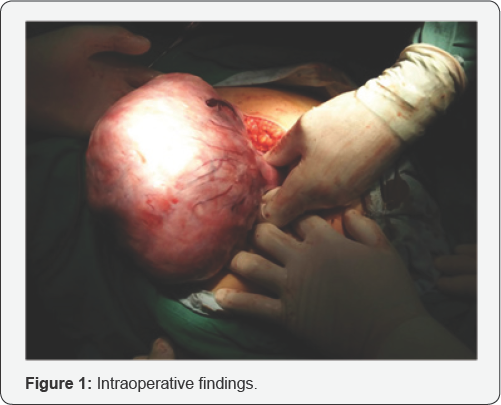
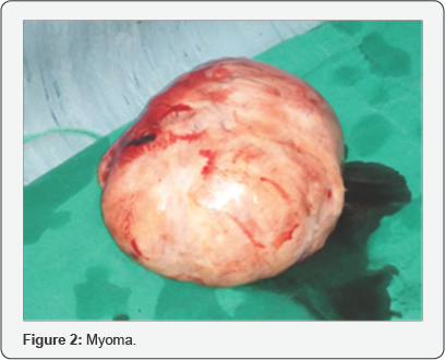
We present a 34 year old nulliparous, Caucasian woman at 14th week of gestation. The patient was admitted in our clinic with a 20cm myoma. A few months ago, before the conception, through ultrasound observation a normal gynecologycal status was found. At 5th gestational week a normal pregnancy was detected, without any signs of myoma. At 7th week of pregnancy a 3.5cm. subserous myoma was found. At 12th week of pregnancy the ultrasound observation showed a subserous pediculated myoma on the left edge of the uterine fundus with dimensions 15/10.9cm. The analysis of the ultrasound observations showed unexpected progressive increasing of the myoma size. Because of the rapid growth and the big size a median laparotomy with myomectomy was performed. A 23 cm pedunculated subserosal myoma with an implantation base of 4cm was found (Figure 1 & 2). The operation and the postoperative period take place without any complications. On the 38th week of pregnancy a healthy, 3400g, full-term, baby boy was delivered by cesarean section. Postpartum and the puerperium period had a regular course.


Discussion
Different outers classify myomas by size in different ways. Most agree that myoma is large when it is over 5cm [11-14]. The Society of Obstetricians and Gynaecologists of Canada, classify myomas over 10cm. like very large [15].
Most ultrasound studies have shown that most of the fibroids during pregnancy (60%-78%) do not show significant changes in their volume. Out of the fibroids that did increase their volume (22% to 32%), the growth was limited in the first trimester [16,17]. There is a prospective study, according to which, if a myoma increases its size during pregnancy, that increase is 12±6% of the initial size, up to a maximum of 25% [16]. A variety of factors, such as genetic and epigenetic mechanism, growth factors, cytokines and disorganization of extracellular matrix components have shown interaction with their pathogenesis [18].
How leiomyomas affect pregnancy is based on their number, size and localization. A significant part of myomas remain asymptomatic and do not require treatment, but 10% lead to complications such as: fetal growth restriction, pelvic pain, placental abruption, placenta previa / accrete, fetal deformities, dystocia, postpartum hemorrhagia [19]. Acceptable indications for myomectomy during pregnancy include intractable pain from a degenerating myoma especially if it is subserosal or pedunculated, a large or rapidly growing myoma, or any large myoma (> 5cm) located in the lower uterine segment [12]. The outcome, both obstetric and neonatal, of women, who underwent myomectomy in pregnancy is comparable to the outcome of those, who were managed conservatively [20,21]. According to few studies, antepartum myomectomy can be safely performed in the first and second trimester of pregnancy, if indicated [20-27].
There is enough scientific data about the safety of laparotomic myomectomy during pregnancy. Recently the number of case reports and studies about the possibility of performing a safe laparoscopic myomectomy during pregnancy is gradually increasing. It begins to slowly replace open surgery because of certain advantages less blood loss, diminished postoperative pain, fewer overall complications, faster recovery and significant cosmetic. However, there are also disadvantages, like the prolonged operative time because of the need of morcellation, especially time-consuming myomas larger than 10cm [16,28].
Morcellation by itself has additional risks of vascular or visceral trauma, and can also lead to leiomyoma dissemination (leiomyomatosis). The possibility of a leiomyosarcoma can't be ruled out, which would lead to a sudden decrease in survivability and upstaging of the disease [16]. Some of those risks can be brought down to a minimum through the use of inbag morcellation, but when a large myoma is present and given the limited area, in cases like ours it is inapplicable. Another disadvantage of the laparoscopic myomectomy during pregnancy is the need of additional training, an experienced surgeon and specialized equipment [16].
Conclusion
Though rarely, rapidly growing and very large myomas can be an indication for myomectomy during the second trimester of pregnancy, because of the limited space for fetus development.
Laparotomic myomectomy should be preferred over laparoscopic myomectomy in cases of very large myomas, due to prolonged operative time, inapplicability of in-bag morcellation and related risks of dissemination of a leiomyoma or leiomyosarcoma.
For more open access journals in JuniperPublishers please click on: https://juniperpublishers.com/
For more articles on Gynecology and Women’s Health please click on: https://juniperpublishers.com/jgwh/
To read more......Fulltext in Gynecology and Women’s Health in Juniper Publishers
https://juniperpublishers.business.site/


Comments
Post a Comment