Juniper Publishers : Ruptured Dermoid Cyst during Pregnancy: A Rare Case Report
JUNIPER PUBLISHERS- JOURNAL OF GYNECOLOGY AND WOMEN’S HEALTH
Journal of Gynecology and Women’s Health-Juniper Publishers
Authored by Aditi Jindal*
Abstract
Aim:We are presenting a rare case report of ruptured mature cystic teratoma during pregnancy.
Background: Mature cystic tutors arise from all the germ cell layers. They have a low malignant potential. They are the most common ovarian tumors presenting in the antenatal period, usually present in the second trimester of pregnancy
Case description:We hereby present a rare case report of ruptured mature cystic teratoma during pregnancy. She was an unbooked patient who presented to us for the first time at 24 weeks with pain abdomen with previous one lower segment caesarean section. She was posted for emergency laparotomy with the suspicion of ovarian cyst torsion . Intraoperatively there was ruptured dermoid cyst in the left ovary. Oopherectomy was performed. She had an uneventful postoperative period.
Conclusion:The most common complication of mature cystic teratoma during pregnancy is torsion. Rupture of a mature cystic teratoma is a rare complication with the reported incidence of 0.2-0.5%.
Clinical Significance:Rupture of dermoid cyst is a rare complication but usually requires surgical intervention.
Keywords: Ruptured dermoid cyst; Pregnancy; Chemical peritonitis
Background
Mature cystic teratoma are cystic tumors derived from the germ cell layers [1]. It typically contains mature tissues of ectodermal (skin, brain), mesodermal (muscle, fat), and endodermal (mucinous or ciliated epithelium) origin. Mature cystic teratoma is the most common benign ovarian tumor in the reproductive age group. It accounts for 20 percent of adult ovarian tumor [2]. The incidence of rupture of mature cystic teratoma varies between 0.2 to 2.5% and the typical presentation is insidious with granulomatous peritonitis. There have been only a few descriptions about rupture of ovarian dermoid tumor during pregnancy in literature.
Case Description
An unbooked 28 year old second gravida, at 24 weeks pregnancy, with previous lower segment caesarean section, presented to us with complaints of pain abdomen since last two days. She had no antenatal visits during pregnancy till now. On general physical examination patient had tachycardia. On abdominal examination there was tenderness on deep palpation however no guarding or rigidity was appreciated. Height of uterus was corresponding to the period of gestation with no suprapubic tenderness. On per vaginal examination os was closed and there was tenderness in the left fornix. An emergency ultrasound was done which revealed a single live intrauterine fetus with a 5x6cm left adenexal mass which showed no vascularity on doppler. With the suspicion of ovarian cyst torsion, patient was posted for emergency laprotomy.

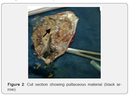
Under spinal anaesthesia, abdomen was opened by a midline vertical scar. Intraoperatively there was cheesy material all over the abdomen densely adherent to the uterus and the gut. Gut was explored for any enteric pathology. Bilateral ovaries were visualised. There was a 4x5cm ruptured ovarian cyst in the left ovary (Figure 1). Decision of oophorectomy was taken and peritoneal lavage done. On gross examination, the cyst contained pultaceous material with hair (Figure 2) Specimen was sent for histopathological examination (HPE). Her postoperative period was uneventful and patient was discharged in satisfactory condition on the 7th post operative day. The HPE report confirmed the diagnosis of dermoid cyst.

Discussion
Mature cystic teratoma usually presents in the reproductive age group. It is usually asymptomatic and diagnosed on routine ultrasonographic examination. The most common complication of mature cystic teratoma is torsion. There have been rare instances of rupture, malignant transformations.
The incidence of rupture is 0.2 to 2.5%. The rare incidence of rupture is attributed to the thick capsule. Dermoid cyst rupture can be primary or secondary. Primary rupture usually occur after torsion, infarction, infection, malignancy or following prolonged pressure due to pregnancy or delivery. Secondary rupture of dermoid cyst may be iatrogenic or following vigorous exercise. Rupture of the teratoma results in leakage of sebaceous material causing an aseptic inflammatory peritoneal reaction leading to chemical peritonitis. The presentation of ruptured dermoid cyst may be acute or chronic. In acute presentation the patient may present as acute abdomen. A similar case has been reported by Chang Y et al. [1] where patient presented after vigorous physical activity. In chronic presentation the patient may present as chronic granulomatous peritonitis, there may be omental deposits mimicking ovarian carcinoma [3,4]. There have been case reports of dermoid cyst with gliomatosis peritonei [5].
Dermoid cyst may remain asymptomatic during pregnancy, and if symptomatic usually has symptoms in the second trimester. The most common pregnancy related complication of dermoid cyst is torsion. There have been few cases of dermoid cyst rupture during pregnancy and at the time of delivery. Maiti S et al. [4] described a similar case of dermoid cyst rupture during pregnancy in early third trimester at 31 weeks where as Nader R et al described rupture of dermoid cyst after delivery [6].
A mature cystic teratoma can be diagnosed at ultrasonography by diffusely or partially echogenic mass with the echogenic area owing to sebaceous material and hair within the cavity. There may be free fluid levels resulting from sebum floating above aqueous fluid, which appears more echogenic than the sebum layer. The diagnosis of mature cystic teratoma at computed tomography (CT) or magnetic resonance imaging (MRI) is fairly straight forward [7]. Usually an asymptomatic dermoid cyst may not require any intervention in less than 6cm tumors [8]. Ruptured dermoid cyst usually require surgical intervention.
Here we have described a rare case of ruptured dermoid cyst during pregnancy.
Clinical Significance
a) Rupture of dermoid cyst is a rare complication but usually requires surgical intervention.
b) Dermoid cyst is the most common ovarian germ cell tumor during pregnancy.
c) Ruptured dermoid cyst may mimic ovarian malignancy.
d) Usually can be diagnosed by USG, MR or CT.
Declaration
Contributors: HC, AJ and PS were involved in the patient management. HC wrote the manuscript. PS contributed in the literature search and corrections in the manuscript. All authors have read and approved the final version of the manuscript.
Contributors: HC, AJ and PS were involved in the patient management. HC wrote the manuscript. PS contributed in the literature search and corrections in the manuscript. All authors have read and approved the final version of the manuscript.
For more open access journals in JuniperPublishers please click on: https://juniperpublishers.com/
For more articles on Gynecology and Women’s Health please click on: https://juniperpublishers.com/jgwh/
To read more......Fulltext in Gynecology and Women’s Health in Juniper Publishers
https://juniperpublishers.business.site/


Comments
Post a Comment